Abstract: "Calcium signaling" contains a unique selection of chapters that cover a wide range of contemporary topics in this ubiquitous and diverse system of cell signaling. This book has the flavor of a primary text book, but it is much more than that. It covers topics ranging from the fundamental aspects of calcium signaling to its clinical implications, in a thoughtful and comprehensive way. It discusses cutting edge researches, and critical issues at depth, and it presents many testable hypotheses for future research. It includes the theoretical and the methodological topics as well as topics related to mathematical modeling, and simulations. If you want to read about calcium signaling in different mammalian cells, oocytes, Zebrafishes, and even in plants, in one and the same book, then this book will not disappoint you. From the beginners to the experts in the field of calcium signaling, everybody will find something useful in this very timely book.
Notes: 68 chapters, 199 illustrations, 143 in color
Abstract: Transient Receptor Potential Channels offers a unique blend of thoughtfully selected topics ranging from the structural biology of this fascinating group of ion channels to their emerging roles in human diseases. This single book covers TRP channels of yeasts, flies, fishes frogs and humans. And from the biophysics of primary thermo-sensory events in cells to the thermosensation at whole organism level, from physiology of pain to the development of pain-killers, from psychiatric illnesses to cancers, from skin cells to sperms, from taste buds to testes, from established facts to heated debates, this book contains something for every TRP enthusiasts, beginner and expert alike. It includes crucial background information, critical analysis of cutting edge research, and ideas and thoughts for numerous testable hypotheses. It also shows directions for future research in this highly dynamic field. It is a book readers will be just as eager to give to others as keep for themselves.
Abstract: This book is a unique and thoughtful blend of critical background information and advances made in a multitude of areas of contemporary islet research. It is an essential reference book, the first of its kind in many years, for professionals as well as for the beginners interested in the study of islet physiology and diabetes. The book is unique in its breadth: it deals with anatomy, histology, ultra-structure, evolution and comparative anatomy, imaging, developmental biology, programming, apoptosis, mitochondrial function, metabolism, cellular signaling, electrophysiology, oscillation of hormone secretion, islets of model animals, immunology, proteomics, regenerative medicine, clinical advances, islet transplantation, and finally islet tumors. Individual chapters contributed by over a hundred experts and enthusiasts, not only provide a balanced view of the recent advances made in the respective fields, but also provide directions and thoughts for future research. Thanks to vivid and colorful illustrations, tables and sketches, the book as a whole, and the individual chapters make reading a pleasant experience. It is a valuable compilation that one would love to possess personally and buy as a present to a colleague.
Notes: Review
"It looks very comprehensive and covers all current areas of interest and includes chapters written by world renowned contributors - very nice!"
Matthias von Herrath, MD
Professor and Director, Center for Type 1 Diabetes Research
La Jolla Institute for Allergy and Immunology,
La Jolla CA, USA
"This book will be welcomed by islet researchers and clinicians interested in the latest developments and theories from which new therapeutics for diabetes can emerge".
Martha Pavlakis, MD
Associate Professor of Medicine, Harvard Medical School
Medical Director, Kidney and Pancreas Transplantation
Beth Israel Deaconess Medical Center
Boston, MA, USA
"The field of islet biology is assuming increasing importance with the recognition that beta-cell dysfunction is a critical feature of type II diabetes. Improving b-cell function and/or b-cell mass may be the best approach to treating diabetes. Thus, the publication of a book covering so many aspects of this field is timely".
Fred Levine, M.D., Ph.D.
Professor, Burnham Institute for Medical Research
Director, Sanford Children's Health Research Center
La Jolla, CA, USA
"The growing prevalence of both type 1 and type 2 diabetes make islet cell biology a critical concern for a wide range of scientists and health professionals. From anatomy and developmental biology, through islet physiology and biochemistry, to issues of immunology and transplantation, this book is a terrific resource".
Jonathan M.W.Slack, Ph.D., F.Med.Sci.
Director, Stem Cell Institute, University of Minnesota,
Medical School
Minneapolis, MN, USA
"This book allows you to enjoy an adventurous and instructive guided tour in the archipelago of the islets of Langerhans. A team of expert and enthusiastic explorers will not only conduct the voyager through almost all niches of this complex and fascinating environment, but also provide him with clues and keys for future exploration of still hidden sites".
Prof. Pellegrino Masiello
Dept. of Experimental Pathology,
University of Pisa,
Pisa, Italy
"The pancreatic islet is a complex mini-organ whose dysfunction is critical in all forms of diabetes. This book provides a comprehensive review of the molecular and cellular processes in islet development, gene expression, and physiology and reviews how a better understanding of the pancreatic islet will advance efforts to develop new therapeutic approaches for diabetes. This book should be quite useful for anyone interested in the pancreatic islet or diabetes".
Alvin C. Powers, M.D.
Joe C. Davis Chair in Biomedical Science
Professor of Medicine, Molecular Physiology and Biophysics
Director, Vanderbilt Diabetes Center
Vanderbilt University
Nashville, TN
USA
"A very timely book on the Islet of Langerhans that coincides with the realization that pancreatic �-cell failure is key to all forms of diabetes mellitus and research in the field is exploding. It contains very thoughtful chapters, each by renown experts in their field, and tackles the complexity of this micro-organ in health and disease very comprehensively and thoughtfully. It will likely become a Reference for years to come for basic researchers, clinicians and students interested is diabetes research"
Marc Prentki, PhD
Professor of Nutrition and Biochemistry
Canada Research Chair in Diabetes and Metabolism
MDRC Director
Montreal, QC
Canada
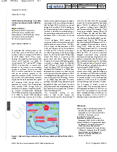
Abstract: Aims: Coiled coil domain containing protein 116 (CCDC116) is a product of the gene coiled coil domain containing 116 located on human chromosome 22. Its function has not yet been established. The present study focuses on the expression of this protein in human pancreatic islets and in the endocrine pancreatic tumors (EPTs). Methods and Results: Expression of the protein was evaluated by immunohistochemistry in endocrine pancreas from six patients and in various EPTs from 51 patients. In pancreatic islets, virtually all insulin, approx. 75% of the somatostatin, and approx. 60% of the pancreatic polypeptide (PP) cells were immunoreactive for the CCDC116 protein whereas glucagon, ghrelin and the exocrine cells were not. All insulinomas, gastrinomas, non-functioning sporadic tumors and the hereditary multihormonal EPTs were immunoreactive with variable relative incidence. Two of the three somatostatinomas, and one of the three ACTH-secreting tumors also expressed CCDC116. Conclusions: The CCDC116 protein is expressed in all islet cell types except the glucagon and ghrelin cells. Most of the EPTs also contained CCDC116 protein. These findings suggest that this protein may play some role for the above mentioned endocrine cells and tumors. Its function has to be investigated in future studies.
Abstract: We have studied whether functional TRPV1 channels exist in the INS-1E cells, a cell type used as a model for b-cells, and
in primary b-cells from rat and human. The effects of the TRPV1 agonists capsaicin and AM404 on the intracellular free
Ca2+ concentration ([Ca2+]i) in the INS-1E cells were studied by fura-2 based microfluorometry. Capsaicin increased [Ca2+]i in a concentration-dependent manner, and the [Ca2+]i increase was dependent on extracellular Ca2+. AM404 also
increased [Ca2+]i in the INS-1E cells. Capsazepine, a specific antagonist of TRPV1, completely blocked the capsaicin- and AM404-induced [Ca2+]i increases. Capsaicin did not increase [Ca2+]i in primary b-cells from rat and human. Whole cell patch clamp configuration was used to record currents across the plasma membrane in the INS-1E cells. Capsaicin
elicited inward currents that were inhibited by capsazepine. Western blot analysis detected TRPV1 proteins in the INS-1E
cells and the human islets. Immunohistochemistry was used to study the expression of TRPV1, but no TRPV1 protein
immunoreactivity was detected in the human islet cells and the human insulinoma cells. We conclude that the INS-1E
cells, but not the primary b-cells, express functional TRPV1 channels.
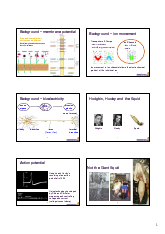
Abstract: In the normal human body pancreatic β-cells spend most of the time in a READY mode rather than in an OFF mode. When in the READY mode, normal β-cells can be easily SWITCHED ON by a variety of apparently trivial stimuli. In the READY mode β-cells are highly excitable because of their high input resistance. A variety of small depolarizing currents mediated through a variety of cation channels triggered by a variety of chemical and physical stimuli can SWITCH ON the cells. Several polymodal ion channels belonging to the transient receptor potential (TRP) family may mediate the depolarizing currents necessary to shift the β-cells from the READY mode to the ON mode. Thanks to the TRP channels, we now know that the Ca2+-activated monovalent cation selective channel described by Sturgess et al. in 1986 (FEBS Lett 208:397�400) is TRPM4, and that the H2O2-activate non-selective cation channel described by Herson and Ashford, in 1997 (J Physiol 501:59�66) is TRPM2. Glucose metabolism generates heat which appears to be a second messenger sensed by the temperature-sensitive TRP channels like the TRPM2 channel. Global knock-out of TRPM5 channel impairs insulin secretion in mice. Other TRPs that may be involved in the regulation of β-cell function include TRPC1, TRPC4, TRPM3, TRPV2 and TRPV4. Future research needs to be intensified to study the molecular regulation of the TRP channels of islets, and to elucidate their roles in the regulation of human β-cell function, in the context of pathogenesis of human islet failure.
Abstract: During a conference titled �TRP channels, from sensory signaling to human disease�, held at the Karolinska Institute, Stockholm, Sweden, on 26th and 27th September, 2009, I was contacted by Springer to publish the proceedings of the
conference. After some discussion with some of the speakers, I understood that that was not going to happen. In stead, we were happy to publish a short meeting report [1]. I thought, the excitement and the momentum that resulted from the
conference could be utilized in compiling a substantial book rather than a modest conference proceeding. The idea for a TRP book appeared very timely.
Abstract: The mechanism by which extracellular ADP ribose (ADPr) increases intracellular free Ca2+ concentration ([Ca2+]i) remains unknown. We measured [Ca2+]i changes in fura-2 loaded rat insulinoma INS-1E cells, and in primary β-cells from rat and human. A phosphonate analogue of ADPr (PADPr) and 8-Bromo-ADPr (8Br-ADPr) were synthesized. ADPr increased [Ca2+]i in the form of a peak followed by a plateau dependent on extracellular Ca2+. NAD+, cADPr, PADPr, 8Br-ADPr or breakdown products of ADPr did not increase [Ca2+]i. The ADPr-induced [Ca2+]i increase was not affected by inhibitors of TRPM2, but was abolished by thapsigargin and inhibited when phospholipase C and IP3 receptors were inhibited. MRS 2179 and MRS 2279, specific inhibitors of the purinergic receptor P2Y1, completely blocked the ADPr-induced [Ca2+]i increase. ADPr increased [Ca2+]i in transfected human astrocytoma cells (1321N1) that express P2Y1 human receptors, but not in untransfected astrocytoma cells. We conclude that ADPr is a specific agonist of P2Y1 receptors.
Abstract: More than 150 participants from 25 countries gathered in Stockholm during 25(th) to 27(th) Sept 2009 to attend the meeting "TRP channels: from sensory signaling to human disease" and enjoyed an international, intensive and vibrant meeting. This meeting shed lights on the recent advances made in this field of research in different sectors of biology, and identified directions for future research and the areas where TRP channels could be used as potential targets for prevention and treatment of human diseases. The participants of this meeting shared their recent largely unpublished data, state-of-the-art techniques and their critical views which would push research in this field forward in the new decade. Another major outcome of this meeting was the realization that extensive work remains to be done to develop the necessary tools and enhance the quality of research in this area so that the prevailing controversies can be resolved. In this report we summarize the latest scientific excitements, some critical issues, as well as some future directions for research that were addressed and discussed in this meeting.
Abstract: Easy access to rodent islets and insulinoma cells and the ease of measuring Ca(2+) by fluorescent indicators have resulted in an overflow of data that have clarified minute details of Ca(2+) signaling in the rodent islets. Our understanding of the mechanisms and the roles of Ca(2+) signaling in the human islets, under physiological conditions, has been hugely influenced by uncritical extrapolation of the rodent data obtained under suboptimal experimental conditions. More recently, electrophysiological and Ca(2+) studies have elucidated the ion channel repertoire relevant for Ca(2+) signaling in the human islets and have examined their relative importance. Many new channels belonging to the transient receptor potential (TRP) family are present in the beta-cells. Ryanodine receptors, nicotinic acid adenine dinucleotide phosphate channel, and Ca(2+)-induced Ca(2+) release add new dimension to the complexity of Ca(2+) signaling in the human beta-cells. A lot more needs to be learnt about the roles of these new channels and CICR, not because that will be easy but because that will be difficult. Much de-learning will also be needed. Human beta-cells do not have a resting state in the normal human body even under physiological fasting conditions. Their membrane potential under physiologically relevant resting conditions is ~-50 mV. Biphasic insulin secretion is an experimental epiphenomenon unrelated to the physiological pulsatile insulin secretion into the portal vein in the human body. Human islets show a wide variety of electrical activities and patterns of [Ca(2+)](i) changes, whose roles in mediating pulsatile secretion of insulin into the portal vein remain questionable. Future studies will hopefully be directed toward a better understanding of Ca(2+) signaling in the human islets in the context of the pathogenesis and treatment of human diabetes.
Abstract: Type 2 melastatin-related transient receptor potential channel (TRPM2), a member of the melastatin-related TRP (transient receptor potential) subfamily is a Ca(2+)-permeable channel activated by hydrogen peroxide (H(2)O(2)). We have investigated the role of TRPM2 channels in mediating the H(2)O(2)-induced increase in the cytoplasmic free Ca(2+) concentration ([Ca(2+)](i)) in insulin-secreting cells. In fura-2 loaded INS-1E cells, a widely used model of beta-cells, and in human beta-cells, H(2)O(2) increased [Ca(2+)](i), in the presence of 3 mM glucose, by inducing Ca(2+) influx across the plasma membrane. H(2)O(2)-induced Ca(2+) influx was not blocked by nimodipine, a blocker of the L-type voltage-gated Ca(2+) channels nor by 2-aminoethoxydiphenyl borate, a blocker of several TRP channels and store-operated channels, but it was completely blocked by N-(p-amylcinnamoyl)anthranilic acid (ACA), a potent inhibitor of TRPM2. Adenosine diphosphate phosphate ribose, a specific activator of TRPM2 channel and H(2)O(2), induced inward cation currents that were blocked by ACA. Western blot using antibodies directed to the epitopes on the N-terminal and on the C-terminal parts of TRPM2 identified the full length TRPM2 (TRPM2-L), and the C-terminally truncated TRPM2 (TRPM2-S) in human islets. We conclude that functional TRPM2 channels mediate H(2)O(2)-induced Ca(2+) entry in beta-cells, a process potently inhibited by ACA.
Abstract: Pancreatic beta-cells have ryanodine receptors but little is known about their physiological regulation. Previous studies have shown that arachidonic acid releases Ca(2+) from intracellular stores in beta-cells but the identity of the channels involved in the Ca(2+) release has not been elucidated. We studied the mechanism by which arachidonic acid induces Ca(2+) concentration changes in pancreatic beta-cells. Cytosolic free Ca(2+) concentration was measured in fura-2-loaded INS-1E cells and in primary beta-cells from Wistar rats. The increase of cytosolic Ca(2+) concentration induced by arachidonic acid (150microM) was due to both Ca(2+) release from intracellular stores and influx of Ca(2+) from extracellular medium. 5,8,11,14-Eicosatetraynoic acid, a non-metabolizable analogue of arachidonic acid, mimicked the effect of arachidonic acid, indicating that arachidonic acid itself mediated Ca(2+) increase. The Ca(2+) release induced by arachidonic acid was from the endoplasmic reticulum since it was blocked by thapsigargin. 2-Aminoethyl diphenylborinate (50microM), which is known to inhibit 1,4,5-inositol-triphosphate-receptors, did not block Ca(2+) release by arachidonic acid. However, ryanodine (100microM), a blocker of ryanodine receptors, abolished the effect of arachidonic acid on Ca(2+) release in both types of cells. These observations indicate that arachidonic acid is a physiological activator of ryanodine receptors in beta-cells.
Abstract: Type 2 diabetes is a polygenic disorder characterized by increased insulin resistance, and impaired insulin secretion leading to abnormalities of glucose and lipid metabolism. Reduced responsiveness of the beta-cells to glucose is a critical feature of this syndrome. Glucagon-like peptide 1, a product of the pro-glucagon gene makes beta-cells competent and has many other anti-diabetic properties. We speculated whether GLP-1-based gene therapy could be an approach for treatment of type 2 diabetes. We started with a clone of rat insulinoma cells (S4 cells), which showed reduced responsiveness to glucose in terms of insulin secretion. We transfected these cells with a plasmid encoding a mutated form of GLP-1 (GLP-1-Gly8), which is resistant to the degrading enzyme dipeptidyl-peptidase IV. Activity of secreted GLP-1-Gly8 was assayed using Chinese hamster lung fibroblasts (CHL) cells that expressed cloned GLP-1 receptor and that were transfected with CRE-Luc. Stable cell lines (Glipsulin cells) obtained by this means produced and stored immunoreactive GLP-1-Gly8. In addition to insulin, the Glipsulin cells secreted the GLP-1-Gly8. The secreted GLP-1-Gly8 was active as evidenced by the ability of the conditioned media to elevate cAMP levels in CHL cells expressing GLP-1 receptors. Glipsulin cells responded to glucose with a 6.8 fold increase in insulin secretion compared to a 2.2 fold increase in the control cells. Our results demonstrate that prolonged exposure to GLP-1-Gly8 secreted by increases glucose-responsiveness of these cells. We speculate that engineering GLP-1-Gly8 secretion by beta-cells is a potential gene therapeutic strategy to treat diabetes.
Abstract: A large number of ion channels, enzymes, pumps and binding proteins participate in the generation of intracellular Ca2+ signals and their decoding. Ca2+ signalling takes place in the form of oscillations, waves and sparks. Such Ca2+ signals occur in almost all cells and regulate diverse cell functions. Perturbation of Ca2+ signalling leads to disease states. Drugs that act on Ca2+-signalling are commonly used in treatment of several diseases. Different molecules involved in Ca2+ signalling are potential targets for development of new therapies. Thus, basic research in Ca2+ signalling increases our understanding of the pathogenesis of diseases at a molecular level and the likelihood of development of new therapeutic modalities.
Abstract: Galanin is a neurotransmitter peptide that suppresses insulin secretion. The present study aimed at investigating how a non-peptide galanin receptor agonist, galnon, affects insulin secretion from isolated pancreatic islets of healthy Wistar and diabetic Goto-Kakizaki (GK) rats. Galnon stimulated insulin release potently in isolated Wistar rat islets; 100 microM of the compound increased the release 8.5 times (p<0.001) at 3.3 mM and 3.7 times (p<0.001) at 16.7 mM glucose. Also in islet perifusions, galnon augmented several-fold both acute and late phases of insulin response to glucose. Furthermore, galnon stimulated insulin release in GK rat islets. These effects were not inhibited by the presence of galanin or the galanin receptor antagonist M35. The stimulatory effects of galnon were partly inhibited by the PKA and PKC inhibitors, H-89 and calphostin C, respectively, at 16.7 but not 3.3 mM glucose. In both Wistar and GK rat islets, insulin release was stimulated by depolarization of 30 mM KCl, and 100 microM galnon further enhanced insulin release 1.5-2 times (p<0.05). Cytosolic calcium levels, determined by fura-2, were increased in parallel with insulin release, and the L-type Ca2+-channel blocker nimodipine suppressed insulin response to glucose and galnon. In conclusion, galnon stimulates insulin release in islets of healthy rats and diabetic GK rats. The mechanism of this stimulatory effect does not involve galanin receptors. Galnon-induced insulin release is not glucose-dependent and appears to involve opening of L-type Ca2+-channels, but the main effect of galnon seems to be exerted at a step distal to these channels, i.e., at B-cell exocytosis.
Abstract: There is little information available concerning the link between the ryanodine (RY) receptors and the downstream Ca(2+) signaling events in beta-cells. In fura-2 loaded INS-1E cells, activation of RY receptors by 9-methyl 5,7-dibromoeudistomin D (MBED) caused a rapid rise of [Ca(2+)]i followed by a plateau and repetitive [Ca(2+)]i spikes on the plateau. The [Ca(2+)]i plateau was abolished by omission of extracellular Ca(2+) and by SKF 96365. In the presence of SKF 96365, MBED produced a transient increase of [Ca(2+)]i, which was abolished by thapsigargin. Activation of RY receptors caused Ca(2+) entry even when the ER Ca(2+) pool was depleted by thapsigargin. The [Ca(2+)]i plateau was not inhibited by nimodipine or ruthenium red, but was inhibited by membrane depolarization, La(3+), Gd(3+), niflumic acid, and 2-aminoethoxydiphenyl borate, agents that inhibit the transient receptor potential channels. The [Ca(2+)]i spikes were inhibited by nimodipine and ryanodine, indicating that they were due to Ca(2+) influx through the voltage-gated Ca(2+) channels and Ca(2+)-induced Ca(2+) release (CICR). Activation of RY receptors depolarized membrane potential as measured by patch clamp. Thus, activation of RY receptors leads to coherent changes in Ca(2+) signaling, which includes activation of TRP-like channels, membrane depolarization, activation of the voltage-gated Ca(2+) channels and CICR.
Abstract: The ryanodine (RY) receptors in beta-cells amplify signals by Ca2+-induced Ca2+ release (CICR). The role of CICR in insulin secretion remains unclear in spite of the fact that caffeine is known to stimulate secretion. This effect of caffeine is attributed solely to the inhibition of cAMP-phosphodiesterases (cAMP-PDEs). We demonstrate that stimulation of insulin secretion by caffeine is due to a sensitization of the RY receptors. The dose-response relationship of caffeine-induced inhibition of cAMP-PDEs was not correlated with the stimulation of insulin secretion. Sensitization of the RY receptors stimulated insulin secretion in a context-dependent manner, that is, only in the presence of a high concentration of glucose. This effect of caffeine depended on an increase in [Ca2+]i. Confocal images of beta-cells demonstrated an increase in [Ca2+]i induced by caffeine but not by forskolin. 9-Methyl-7-bromoeudistomin D (MBED), which sensitizes RY receptors, did not inhibit cAMP-PDEs, but it stimulated secretion in a glucose-dependent manner. The stimulation of secretion by caffeine and MBED involved both the first and the second phases of secretion. We conclude that the RY receptors of beta-cells mediate a distinct glucose-dependent signal for insulin secretion and may be a target for developing drugs that will stimulate insulin secretion only in a glucose-dependent manner.
Abstract: The list of Ca(2+) channels involved in stimulus-secretion coupling in beta-cells is increasing. In this respect the roles of the voltage-gated Ca(2+) channels and IP(3) receptors are well accepted. There is a lack of consensus about the significance of a third group of Ca(2+) channels called ryanodine (RY) receptors. These are large conduits located on Ca(2+) storage organelle. Ca(2+) gates these channels in a concentration- and time-dependent manner. Activation of these channels by Ca(2+) leads to fast release of Ca(2+) from the stores, a process called Ca(2+)-induced Ca(2+) release (CICR). A substantial body of evidence confirms that beta-cells have RY receptors. CICR by RY receptors amplifies Ca(2+) signals. Some properties of RY receptors ensure that this amplification process is engaged in a context-dependent manner. Several endogenous molecules and processes that modulate RY receptors determine the appropriate context. Among these are several glycolytic intermediates, long-chain acyl CoA, ATP, cAMP, cADPR, NO, and high luminal Ca(2+) concentration, and all of these have been shown to sensitize RY receptors to the trigger action of Ca(2+). RY receptors, thus, detect co-incident signals and integrate them. These Ca(2+) channels are targets for the action of cAMP-linked incretin hormones that stimulate glucose-dependent insulin secretion. In beta-cells some RY receptors are located on the secretory vesicles. Thus, despite their low abundance, RY receptors are emerging as distinct players in beta-cell function by virtue of their large conductance, strategic locations, and their ability to amplify Ca(2+) signals in a context-dependent manner.
Abstract: Stimulus-secretion coupling in pancreatic beta-cells involves membrane depolarization and Ca(2+) entry through voltage-gated L-type Ca(2+) channels, which is one determinant of increases in the cytoplasmic free Ca(2+) concentration ([Ca(2+)](i)). We investigated how the endoplasmic reticulum (ER)-associated Ca(2+) apparatus further modifies this Ca(2+) signal. When fura-2-loaded mouse beta-cells were depolarized by KCl in the presence of 3 mm glucose, [Ca(2+)](i) increased to a peak in two phases. The second phase of the [Ca(2+)](i) increase was abolished when ER Ca(2+) stores were depleted by thapsigargin. The steady-state [Ca(2+)](i) measured at 300 s of depolarization was higher in control cells compared with cells in which the ER Ca(2+) pools were depleted. The amount of Ca(2+) presented to the cytoplasm during depolarization as estimated from the integral of the increment in [Ca(2+)](i) over time (integralDelta[Ca(2+)](i).dt) was approximately 30% higher compared with that in the Ca(2+) pool-depleted cells. neo-thapsigargin, an inactive analog, did not affect [Ca(2+)](i) response. Using Sr(2+) in the extracellular medium and exploiting the differences in the fluorescence properties of Ca(2+)- and Sr(2+)-bound fluo-3, we found that the incoming Sr(2+) triggered Ca(2+) release from the ER. Depolarization-induced [Ca(2+)](i) response was not altered by, an inhibitor of phosphatidylinositol-specific phospholipase C, suggesting that stimulation of the enzyme by Ca(2+) is not essential for amplification of Ca(2+) signaling. [Ca(2+)](i) response was enhanced when cells were depolarized in the presence of 3 mm glucose, forskolin, and caffeine, suggesting involvement of ryanodine receptors in the amplification process. Pretreatment with ryanodine (100 microm) diminished the second phase of the depolarization-induced increase in [Ca(2+)](i). We conclude that Ca(2+) entry through L-type voltage-gated Ca(2+) channels triggers Ca(2+) release from the ER and that such a process amplifies depolarization-induced Ca(2+) signaling in beta-cells.
Abstract: OBJECTIVE: Previous studies have demonstrated that leptin can stimulate proliferation of insulin-secreting tumor cell lines. The objective of this study was to characterize whether leptin could stimulate proliferation of primary beta-cells too. Since adult beta-cells have very limited capacity for replication, we examined the effect of leptin on islets of Langerhans obtained from fetal rats, in a tissue culture system. METHODS: Leptin receptor mRNA and c-fos mRNA were measured by RT-PCR. Proliferation of fetal rat islet cells was measured by a WST-1 colorimetric assay and [3H]-thymidine incorporation assay. RESULTS: Leptin stimulated proliferation of serum-deprived fetal rat islet cells, as indicated by increased formation of formazan dye from a tetrazolium salt WST-1. Leptin stimulated DNA synthesis in islet cells, as indicated by increased [3H]-thymidine incorporation into DNA. The effect of leptin on islet cell proliferation was on average 39-50% of the effect obtained with 10% fetal bovine serum. Leptin increased c-fos mRNA expression by 2.8-fold in isolated fetal islets after 30 min treatment. In fetal pancreatic islets, both the common extracellular portion (OB-R) and the intact long form (OB-Rb) of the leptin receptor were readily detected by reverse transcriptase polymerase chain reaction. CONCLUSION: Functional leptin receptors are expressed in pancreatic islet cells, as early as during the fetal stage of development of these microorgans. Leptin stimulates proliferation of fetal islet cells and might play a role in determining islet cell mass at birth.
Abstract: Neisseria gonorrhoeae and Neisseria meningitidis are Gram-negative bacterial pathogens that infect human mucosal epithelia. Type IV pilus-mediated adherence of these bacteria is a crucial early event for establishment of infection. In this work, we show that the type IV pili transduce a signal into the eucaryotic host cell. Purified adherent pili, but not pili from a low binding mutant, trigger an increase in the cytosolic free calcium ([Ca2+]i) in target epithelial cells, a signal known to control many cellular responses. The [Ca2+]i increase was blocked by antibodies against CD46, a putative pilus receptor, suggesting a role for this protein in signal transduction. Pilus-mediated attachment was inhibited by depletion of host cell intracellular Ca2+ stores but not by removal of extracellular Ca2+. Further, kinase inhibition studies showed that pilus-mediated adherence is dependent on casein kinase II. In summary, these data reveal a novel function of the type IV pili, namely induction of signal transduction pathways in host cells.
Abstract: Molecular mechanisms that regulate in situ activation of ryanodine receptors (RY) in different cells are poorly understood. Here we demonstrate that caffeine (10 mM) released Ca2+ from the endoplasmic reticulum (ER) in the form of small spikes in only 14% of cultured fura-2 loaded beta cells from ob/ob mice. Surprisingly, when forskolin, an activator of adenylyl cyclase was present, caffeine induced larger Ca2+ spikes in as many as 60% of the cells. Forskolin or the phosphodiesterase-resistant PKA activator Sp-cAMPS alone did not release Ca2+ from ER. 4-Chloro-3-ethylphenol (4-CEP), an agent that activates RYs in other cell systems, released Ca2+ from ER, giving rise to a slow and small increase in [Ca2+]i in beta cells. Prior exposure of cells to forskolin or caffeine (5 mM) qualitatively altered Ca2+ release by 4-CEP, giving rise to Ca2+ spikes. In glucose-stimulated beta cells forskolin induced Ca2+ spikes that were enhanced by 3,9-dimethylxanthine, an activator of RYs. Analysis of RNA from islets and insulin-secreting betaTC-3-cells by RNase protection assay, using type-specific RY probes, revealed low-level expression of mRNA for the type 2 isoform of the receptor (RY2). We conclude that in situ activation of RY2 in beta cells requires cAMP-dependent phosphorylation, a process that recruits the receptor in a functionally operative form.
Abstract: In single mouse skeletal muscle fibers injected with fluorescent Ca2+ indicator Indo-1, 4-chloro-m-cresol (chlorocresol, 4-CmC) and its lipophilic analogue 4-chloro-3-ethylphenol (4-CEP) increased resting myoplasmic free [Ca2+] ([Ca2+]i) in a dose-dependent manner. In this regard, 4-CEP was more potent than 4-CmC and both were more potent than caffeine. High concentrations of 4-CmC (1 mM) or 4-CEP (500 microM) caused large and irreversible increase in resting [Ca2+]i leading to contracture. 4-CmC potentiated the [Ca2+]i increase and force of contraction induced by tetanic stimulation. Unlike caffeine, 4-CmC did not affect the activity of sarcoplasmic reticulum Ca2+ pump or the myofibrillar Ca2+ sensitivity. A low concentration of 4-CEP (20 microM) had no effect on resting [Ca2+]i on its own, but it enhanced the resting [Ca2+]i increase induced by caffeine and also potentiated the [Ca2+]i increase and contraction induced by tetanic stimulation. However, a relatively high concentration of 4-CEP (200 microM) inhibited tetanic stimulation-induced [Ca2+]i increase and contraction. Dantrolene, a muscle relaxant, inhibited 4-CmC-induced [Ca2+]i increase under resting conditions. However, when 4-CEP was applied in the presence of dantrolene, there was an exaggerated increase in [Ca2+]i. We conclude that 4-CmC and 4-CEP are potent agonists that can increase [Ca2+]i rapidly and reversibly by activating ryanodine receptors in situ in intact skeletal muscle fibers. These compounds, specially 4-CmC, may be useful for mechanistic and functional studies of ryanodine receptors and excitation-contraction coupling in skeletal muscles.
Abstract: In addition to its interaction at hypothalamic sites to affect feeding and energy expenditure, leptin has been shown to exhibit a proliferative response in erythropoietic cells. The functional leptin receptor is also present in pancreatic islets and we now demonstrate that a commonly used clonal insulin secreting beta-cell line, RINm5F, expresses high levels of the Ob-Rb mRNA. Leptin causes an increase in tyrosine phosphorylation of a number of intracellular proteins and a dose related (10 nM-200 nM) increase in expression of the immediate-early gene, c-fos. This precedes a leptin induced proliferative response in serum-deprived RINm5F cells, which suggests that leptin might be involved in the complex regulation of proliferation of the pancreatic beta-cell.
Abstract: 2,2'-Dithiodipyridine (2,2'-DTDP), a reactive disulphide that mobilizes Ca2+ from ryanodine-sensitive Ca2+ stores in muscle, induced a biphasic increase in cytoplasmic free Ca2+ concentration ([Ca2+]i) in pancreatic beta-cells loaded with fura 2. This increase consisted of an early transient followed by a second, slower, rise. The [Ca2+]i transient was dependent on extracellular Ca2+ and disappeared on treatment with nimodipine. The reactive disulphide caused plasma membrane depolarization, as studied by the perforated-patch configuration of the patch-clamp technique. Hence membrane depolarization and opening of the L-type voltage-gated Ca2+ channels were responsible for the first transient in [Ca2+]i. The second slower increase in [Ca2+]i was prolonged but readily reversed by the disulphide-reducing agent 1,4-dithiothreitol. This increase in [Ca2+]i was not decreased by nimodipine or by omission of extracellular Ca2+, but was eliminated when the Ins(1,4,5)P3-sensitive Ca2+ pool was first depleted by carbachol. Ryanodine or its beta-alanyl analogue did not release Ca2+ from intracellular stores, and a high concentration of ryanodine did not inhibit Ca2+ release by 2,2'-DTDP. The disulphide compound suppressed glucose metabolism and decreased the mitochondrial inner-membrane potential. We conclude that thiol oxidation by 2,2'-DTDP affects Ca2+ homeostasis in beta-cells by multiple mechanisms. However, unlike the situation in muscle, in beta-cells 2,2'-DTDP releases Ca2+ from intracellular pools by mechanisms that do not involve activation of ryanodine receptors. Instead, in these cells the Ins(1,4,5)P3-sensitive intracellular Ca2+ store comprises an alternative target for the Ca(2+)-mobilizing action of the reactive disulphide compound.
Abstract: Stimulation of pancreatic beta-cells by glucose gives rise to an increase in the cytoplasmic free calcium concentration ([Ca2+]i) and exocytosis of insulin. Cyclic adenosine 5'-diphosphate ribose (cADPR), a metabolite of beta-NAD+, has been reported to increase [Ca2+]i in pancreatic beta-cells by releasing Ca2+ from inositol 1,4,5-trisphosphate-insensitive intracellular stores. In the present study, we have examined the role of cADPR in glucose-mediated increases in [Ca2+]i and insulin exocytosis. Dispersed ob/ob mouse beta-cell aggregates were either pressure microinjected with fura-2 salt or loaded with fura-2 acetoxymethyl ester, and [Ca2+]i was monitored by microfluorimetry. Microinjection of beta-NAD+ into fura-2-loaded beta-cells did not increase [Ca2+]i nor did it alter the cells' subsequent [Ca2+]i response to glucose. Cells microinjected with the cADPR antagonist 8NH2-cADPR increased [Ca2+]i in response to glucose equally well as those injected with cADPR. Finally, the ability of cADPR to promote exocytosis of insulin in electropermeabilized beta-cells was investigated. cADPR on its own did not increase insulin secretion nor did it potentiate Ca2+-induced insulin secretion. We conclude that cADPR neither plays a significant role in glucose-mediated increases in [Ca2+]i nor interacts directly with the molecular mechanisms regulating exocytosis of insulin in normal pancreatic beta-cells.
Abstract: In the pancreatic beta-cell, an increase in the cytoplasmic free Ca2+ concentration ([Ca2+]i) by caffeine is believed to indicate mobilization of Ca2+ from intracellular stores, through activation of a ryanodine receptor-like channel. It is not known whether other mechanisms, as well, underlie caffeine-induced changes in [Ca2+]i. We studied the effects of caffeine on [Ca2+]i by using dual-wavelength excitation microfluorimetry in fura-2-loaded beta-cells. In the presence of a non-stimulatory concentration of glucose, caffeine (10-50 mM) consistently increased [Ca2+]i. The effect was completely blocked by omission of extracellular Ca2+ and by blockers of the L-type voltage-gated Ca2+ channel, such as D-600 or nifedipine. Depletion of agonist-sensitive intracellular Ca2+ pools by thapsigargin did not inhibit the stimulatory effect of caffeine on [Ca2+]i. Moreover, this effect of caffeine was not due to an increase in cyclic AMP, since forskolin and 3-isobutyl-1-methylxanthine (IBMX) failed to raise [Ca2+]i in unstimulated beta-cells. In beta-cells, glucose and sulphonylureas increase [Ca2+]i by causing closure of ATP-sensitive K+ channels (KATP channels). Caffeine also caused inhibition of KATP channel activity, as measured in excised inside-out patches. Accordingly, caffeine (> 10 mM) induced insulin release from beta-cells in the presence of a non-stimulatory concentration of glucose (3 mM). Hence, membrane depolarization and opening of voltage-gated L-type Ca2+ channels were the underlying mechanisms whereby the xanthine drug increased [Ca2+]i and induced insulin release. Paradoxically, in glucose-stimulated beta-cells, caffeine (> 10 mM) lowered [Ca2+]i. This effect was due to the fact that caffeine reduced depolarization-induced whole-cell Ca2+ current through the L-type voltage-gated Ca2+ channel in a dose-dependent manner. Lower concentrations of caffeine (2.5-5.0 mM), when added after glucose-stimulated increase in [Ca2+]i, induced fast oscillations in [Ca2+]i. The latter effect was likely to be attributable to the cyclic AMP-elevating action of caffeine, leading to phosphorylation of voltage-gated Ca2+ channels. Hence, in beta-cells, caffeine-induced changes in [Ca2+]i are not due to any interaction with intracellular Ca2+ pools. In these cells, a direct interference with KATP channel- and L-type voltage-gated Ca(2+)-channel activity is the underlying mechanism by which caffeine increases or decreases [Ca2+]i.
Abstract: The biological activity of many proteins, including voltage-sensitive ion channels, is controlled by their state of phosphorylation. Ca2+ influx through voltage-activated L-type Ca2+ channels serves as the major stimulatory signal in insulin-secreting cells. We have now investigated the extent to which Ca2+ handling in clonal insulin-secreting RiNm5F cells was affected by okadaic acid, an inhibitor of various serine/threonine protein phosphatases. Whole-cell patch-clamp experiments showed that okadaic acid generated an increase in membrane current, suggesting that it promotes Ca2+ influx through L-type voltage-gated Ca2+ channels probably by modifying their phosphorylation state. Okadaic acid was found to provoke a transient rise in the cytoplasmic free Ca2+ concentration ([Ca2+]i) but had no further effect on the K(+)-induced increase. The Ca2+ transient induced by okadaic acid was dependent on the presence of extracellular Ca2+ and was abolished by D600, a blocker of voltage-activated L-type Ca2+ channels. Concomitant with the rise in [Ca2+]i, okadaic acid induced insulin secretion, a phenomenon that was also dependent on extracellular Ca2+. It is proposed that hyperphosphorylation of voltage-activated L-type Ca2+ channels in insulin-secreting cells lowers the threshold potential for their activation.
Abstract: We characterized and directly compared the Ca(2+)-releasing actions of two inhibitors of endoplasmic-reticulum (ER) Ca(2+)-ATPase, thapsigargin and 2,5-di-(t-butyl)-1,4-benzohydroquinone (tBuBHQ), in electropermeabilized insulin-secreting RINm5F cells. Ambient free calcium concentration ([Ca2+]) was monitored by Ca(2+)-selective mini-electrodes. After ATP-dependent Ca2+ uptake, thapsigargin and tBuBHQ released Ca2+ with and EC50 of approximately 37 nM and approximately 2 microM respectively. Both agents mobilized Ca2+ predominantly from the Ins(1,4,5)P3-sensitive Ca2+ pool, and in this respect thapsigargin was more specific than tBuBHQ. The total increase in [Ca2+] obtained with thapsigargin and Ins(1,4,5)P3 was, on the average, only 7% greater than that with Ins(1,4,5)P3 alone. In contrast, the total increase in [Ca2+] obtained with tBuBHQ and Ins(1,4,5)P3 was 33% greater than that obtained with only InsP3 (P < 0.05). Although Ca2+ was rapidly mobilized by thapsigargin and tBuBHQ, complete depletion of the Ins(1,4,5)P3-sensitive Ca2+ pool was difficult to achieve. After the release by thapsigargin or tBuBHQ, Ins(1,4,5)P3 induced additional Ca2+ release. The additional Ins(1,4,5)P3-induced Ca2+ release was not altered by supramaximal concentrations of thapsigargin and tBuBHQ, or by Bafilomycin A1, an inhibitor of V-type ATPases, but was decreased by prolonged treatment with the ER Ca(2+)-ATPase inhibitors. These results suggest the existence of distinct uptake and release compartments within the Ins(1,4,5)P3-sensitive Ca2+ pool. When treated with the inhibitors, the two compartments became distinguishable on the basis of their Ca2+ permeability. Apparently, thapsigargin and tBuBHQ readily mobilized Ca2+ from the uptake compartment, whereas Ca2+ from the release compartment could be mobilized only very slowly, in the absence of Ins(1,4,5)P3.
Abstract: Effects of sulfhydryl modification on the ATP regulated K+ channel (KATP channel) in the pancreatic beta-cell were studied, using the patch clamp technique. Application of the sulfhydryl oxidizing agents thimerosal and 2,2'-dithio-bis(5-nitropyridine) (DTBNP), in micromolar concentrations, caused complete inhibition of the KATP channel, in inside-out patches. The inhibition was rapid and was reversed by the disulfide reducing agents dithiothreitol and cysteine. Thimerosal, which is poorly membrane permeable, inhibited channel activity, only when applied to the intracellular face of the plasma membrane. In contrast, DTBNP, which is highly lipophilic, caused closure of the KATP channel and consequent depolarization of the membrane potential, also when applied extracellularly. Our results indicate the presence of accessible free SH groups on the cytoplasmic side of the KATP channel in the pancreatic beta-cell. These SH groups are essential for channel function and it is possible that thiol-dependent redox mechanisms can modulate KATP channel activity.
Abstract: The sulphydryl reagent thimerosal (50 microM) released Ca2+ from a non-mitochondrial intracellular Ca2+ pool in a dose-dependent manner in permeabilized insulin-secreting RINm5F cells. This release was reversed after addition of the reducing agent dithiothreitol. Ca2+ was released from an Ins(1,4,5)P3-insensitive pool, since release was observed even after depletion of the Ins(1,4,5)P3-sensitive pool by a supramaximal dose of Ins(2,4,5)P3 or thapsigargin. The Ins(1,4,5)P3-sensitive pool remained essentially unaltered by thimerosal. Thimerosal-induced Ca2+ release was potentiated by caffeine. These findings suggest the existence of Ca(2+)-induced Ca2+ release also in insulin-secreting cells.
Abstract: Electropermeabilised insulin-secreting RINm5F cells sequestered Ca2+, resulting in a steady-state level of the ambient free Ca2+ concentration corresponding to 723 +/- 127 nM (mean +/- SEM, n = 10), as monitored by a Ca(2+)-selective minielectrode. Inositol 1,4,5-trisphosphate (Ins(1,4,5)P3) promoted a rapid and pronounced release of Ca2+. This Ca2+ was resequestered and a new steady-state Ca2+ level was attained, which was always lower (460 +/- 102 nM, n = 10, P less than 0.001) than the steady-state Ca2+ level maintained before the addition of Ins(1,4,5)P3. Whereas the initial reuptake of Ca2+ subsequent to Ins(1,4,5)P3 stimulation was relatively slow, the later part of reuptake was fast as compared to the reuptake phases of a pulse addition of extraneous Ca2+. In the latter case the uptake of Ca2+ resulted in a steady-state level similar to that found in the absence of Ins(1,4,5)P3. Addition of Ins(1,4,5)P3 under this condition resulted in a further Ca2+ uptake and thus a lower steady-state Ca2+ level. Heparin, which binds to the Ins(1,4,5)P3 receptor, also lowered the steady-state free Ca2+ concentration. In contrast to Ins(1,4,5)P3, inositol 1,3,4,5-tetrakisphosphate was without effect on Ca2+ sequestration. These findings are consistent with the presence of a high-affinity Ins(1,4,5)P3 receptor promoting continuous release of Ca2+ under basal conditions and/or the Ins(1,4,5)P3 receptor being actively involved in Ca2+ sequestration.
Abstract: In a prospective study disposable insulin syringes were repeatedly used for injecting insulin to 92 insulin-taking diabetic patients admitted in hospital with various illnesses. One separate syringe was used for each patient; the syringes were not washed or boiled and were not kept in spirit or in refrigerator during reuse. Syringes were flushed with air, needle recapped and replaced into their plastic cover. Number of injections made using one syringe was 2 to 120 (mean 31.3, SEM 2.3). For the patients' total insulin - taking period in hospital one disposable syringe per patient was enough in all but six cases. There were 2792 reinjections and incidence of infection of the injection site was zero.
Abstract: Faecal samples from 63 subjects with self-diagnosis of "chronic dysentery" and 50 control subjects were examined under light microscope. Vegetative form of E.h. was not detected in any of them. E.h. cyst was found in 6.3% of the "chronic dysentery" subjects and in 16% of the control subjects. E. coli and Giardia were also detected less often in the "chronic dysentery" than the control subjects. Lower incidence of protozoa namely E.h. cyst, E. coli cyst or Giardia in the former group was probably due to frequent intake of antiamoebic agents which are also effective against other intestinal protozoa. Incidence of Ascaris, hookworm, and Trichuris was not appreciably different in the two groups. More subjects in the "chronic dysentery" group had normal stool findings (31%) as compared to the control subjects (16%). It has been inferred that E.h. infection is not the cause of symptoms of "chronic dysentery".
Abstract: In a retrospective study carried out in Chittagong Medical College the results of HSC and First-Professional MBBS examination of 725 students were analysed. It was concluded from the analysis that if standard of HSC examination is well maintained the results of the said examination correlates well with students' performance in the First Professional MBBS examination. An analysis of admission into Chittagong Medical College in past 20 academic sessions has been made which shows that an increasing number of students securing high marks in HSC are being admitted into Medical College during recent years. The marking standard of HSC examination has however remained fairly uniform over the years except during early post-liberation period.
Abstract: Islets of Langerhans, named after their discoverer Paul Langerhans, constitute a unique endocrine organ of critical importance in the metabolism of nutrients and energy homeostasis. Individual islets consist of three major types of electrically excitable cells, namely b�cells that secrete insulin, a�cells that secrete glucagon and d�cells that secrete somatostatin. Islets develop from the gut endoderm and a set of transcription factors including PDX1, PAX4 and PAX6, play important roles in determination of the cell types and their functions. These microorgans are coordinated by neural and hormonal networks and secret hormones in an oscillatory manner. Nutrient metabolism and incretin hormones trigger insulin secretion. Important cellular components and messengers that determine insulin secretion include glucose transporters, glucokinase, ATP/ADP�ratio, mitochondrial metabolism, ATP�sensitive potassium channels, cAMP and Ca2+. Because of their importance in the pathogenesis of diabetes and increased interest in islet transplantation during recent years, islets of Langerhans continue to be a field of extensive research throughout the world.
Abstract: Previous studies from our group reported that pancreatic beta-cells express ryanodine receptors (RyRs) that can mediate Ca2+-induced Ca2+ release (CICR). The full consequences of the activation of RyRs on Ca2+ signaling in these cells, however, remained unclear. An important open question was whether activation of the RyRs leads to activation of any Ca2+ channels in the plasma membrane, and thereby depolarizes membrane potential. One main aim of the thesis was to address this question. As a corollary, we have also looked for the existence of functional TRPV1 channels, and have elucidated the molecular mechanisms that underlie the [Ca2+]i-elevating effect of ADP ribose in these cells.
We used methods such as measurement of the [Ca2+]i in single cells loaded with fura-2, patch clamp technique, Western blot analysis, immunohistochemistry, a variety of pharmacological tools, and a series of carefully designed protocols. In most experiments, we used S5 cells, derived from the rat insulinoma cell line INS-1E, but we also used primary beta-cells from mice, rat, and human. Activation of the RyRs by 9-methyl 5,7-dibromoeudistomin D (MBED) increased the [Ca2+]i with an initial peak, followed by a decline to a plateau phase, and regenerative spikes superimposed on the plateau. The initial [Ca2+]i increase was due to the activation of the RyRs in the ER, since it was abolished by thapsigargin, but was present when extracellular Ca2+ was omitted or when Ca2+ entry was blocked by
SKF 96365. The plateau phase was due to Ca2+ entry across the plasma membrane, since it was abolished by
omission of extracellular Ca2+, and blocked by SKF 96365. The plateau phase was not solely dependent on
the filling state of the ER, since it was not abolished by thapsigargin. Inhibition of the voltage-gated Ca2+
channels by nimodipine did not inhibit the plateau phase. Several agents that block TRP channels, e.g. La3+,
Gd3+, niflumic acid, and 2-APB, inhibited the plateau phase. It was also inhibited by membrane
depolarization. We conclude that the plateau phase was due to activation of some TRP-like channels.
Activation of RyRs by MBED also induced membrane depolarization. The spikes required Ca2+ entry
through the L-type voltage-gated Ca2+ channels, as they were abolished by nimodipine. The spikes resulted
from CICR, since they were inhibited in a use-dependent way by ryanodine, and abolished after depletion of
the ER by thapsigargin. Thus, activation of RyRs activated TRP-like channels, depolarized the plasma
membrane, activated L-type voltage-gated Ca2+ channels and triggered CICR.
During the course of this thesis we reported that TRPM2 is present in the INS1-E cells and the
human beta-cells. We studied whether TRPM2 was involved in the Ca2+ entry triggered by the activation of
RyRs. N-(p-amylcinnamoyl) anthranilic acid (ACA), an inhibitor of TRPM2, did not inhibit the MBEDinduced
[Ca2+]i entry. ADP ribose (ADPr), when applied intracellularly, is an agonist of TRPM2. We found
that extracellularly applied ADPr increased [Ca2+]i in the form of an initial peak followed by a plateau that
depended on extracellular Ca2+. EC50 of ADPr was ~30 μM. NAD+, cADPr, a phosphonate analogue of
ADPr (PADPr), 8-bromo-ADPr or breakdown products of ADPr did not increase [Ca2+]i. Inhibitors of
TRPM2, e.g. flufenamic acid, niflumic acid, and ACA did not affect the ADPr-induced [Ca2+]i increase.
Two specific inhibitors of the purinergic receptor P2Y1, e.g. MRS 2179 and MRS 2279 completely blocked
the ADPr-induced [Ca2+]i increase. The [Ca2+]i increase by ADPr required activation of PI-PLC, since the
PI-PLC inhibitor U73122 abolished the [Ca2+]i increase. The ADPr-induced [Ca2+]i increase was through the
IP3 receptors, since it was inhibited by 2-APB, an inhibitor of the IP3 receptors. ADPr increased [Ca2+]i in the
transfected human astrocytoma cells that expressed the P2Y1 receptors, but not in the wild type astrocytoma
cells. We conclude that extracellular ADPr is an endogenous and specific agonist of P2Y1 receptors.
Capsaicin and AM404, two specific agonists of TRPV1, increased [Ca2+]i in the INS-1E cells.
Capsazepine, a specific antagonist of TRPV1, completely blocked the capsaicin-induced [Ca2+]i increase.
Capsaicin elicited inward currents that were abolished by capsazepine. TRPV1 protein was detected in the
INS-1E cells and human beta-cells by Western blot. However, no TRPV1 immunoreactivity was detected in
the human islet cells and human insulinoma by immunohistochemistry. Capsaicin did not increase [Ca2+]i in
primary beta-cells from rat or human. We conclude that INS-1E cells express functional TRPV1 channels.
In summary, we have shown that (1) RyR activation leads to activation of TRP-like channels in the
plasma membrane, membrane depolarization, activation of L-type voltage-gated Ca2+ channels and CICR.
(2) ADPr is a specific and endogenous low affinity ligand for the P2Y1 receptors. (3) Functional TRPV1 channels are expressed in the INS-1E cells, but not in the primary beta-cells.
Abstract: FKBP12 is a 11.8-kDa immunophilin to which the immunosuppressant drugs FK506 (tacrolimus) and rapamycin (sirolimus) bind with high affinity. Like many of these immunophilins, FKBP12 possesses cis�trans peptidyl-prolyl isomerase or rotamase activity. This enzymatic activity is thought to accelerate the rate at which proteins fold into their native conformation. However, inhibition of the activity is not responsible for immunosuppressive action of the drugs that bind to FKBP12. The cellular role of this enzymatic function has not been clearly defined. FKBP12 is required for T-cell receptor-mediated signal transduction that leads to transcription of the T-cell growth factor interleukin-2. Calcineurin is a common target of the FKBP�FK506 complex. FKBP12 and its homolog FKBP12.6 modulate the calcium release activity of skeletal and cardiac ryanodine receptors, respectively. FKBP12 and FKBP12.6 are also known as calstablin 1 or 2 respectively. The immunosuppressive drug FK506 (tacrolimus) binds tightly to FKBP12 and inhibits its cis�trans peptidyl-prolyl isomerase activity. The FK506�FKBP12 complex inhibits calcineurin and thereby inhibits the transcription of early T-cell activation genes.